Core Instrumentation Facility (CIF)
The core instrumentation facility – A central facility dedicated exclusively for machines. We have wide range of machines in the CIF to facilitate cutting edge research. The facility is designed in a way that anyone from the institute can access these instruments according to their convenience. The Core instrumentation facility will provide required training and support to the machine users. The users from other institutes can also make use of the facilities.
Other instruments in the facility includes
Multifunctinal Electro Fusion & Electroporation Unit with Hybridoma Production
Make
BTX Harvard Apparatus
Model
ECM-2001 LITE
Ultra-Centrifugation facility
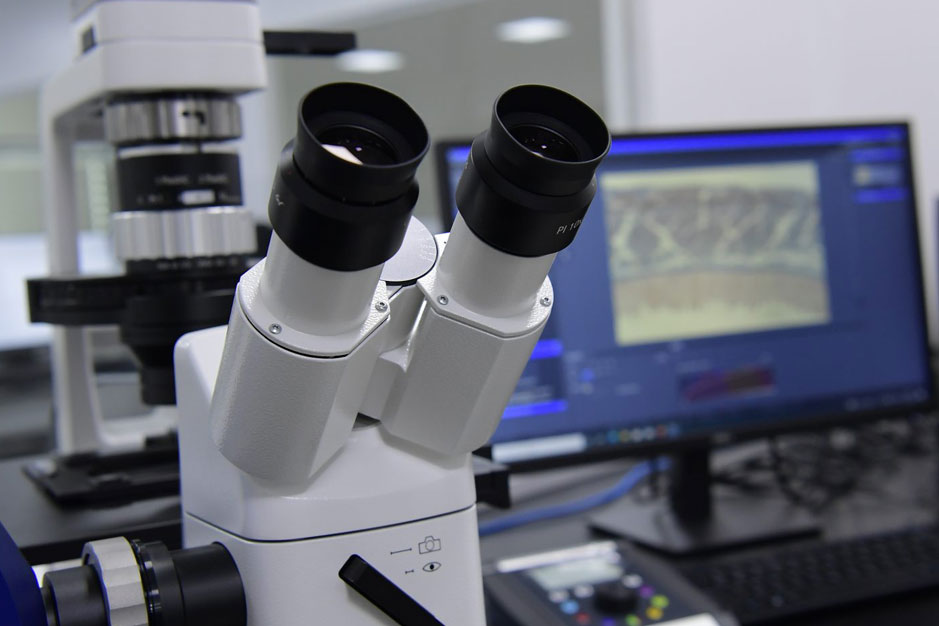
Confocal laser scanning microscopy
Fast protein liquid chromatography
Fast protein liquid chromatography (FPLC) is a form of medium pressure chromatography originally developed for purifying proteins with high resolution and reproducibility. Its distinguishing feature is that the stationary phase is composed of small-diameter beads (generally cross-linked agarose) that are packed in glass or plastic columns and have high loading capacity. Resins for FPLC are available in a wide range of particle sizes and ligand surfaces, which are selected on the basis of their application. The FPLC system allows the use of a wide range of aqueous buffers (the mobile phase) and different stationary phases to perform the main chromatography modes (ion exchange, gel filtration, affinity, chromatofocusing, hydrophobic interaction, reverse phase). However, anion exchange and gel filtration chromatography are the modes most commonly used.
Fluorescence-activated cell sorting
Fluorescence-activated cell sorting (FACS) is a technique to purify specific cell populations based on phenotypes detected by flow cytometry. This method enables researchers to better understand the characteristics of a single-cell population without the influence of other cells. Compared to other methods of cell enrichment, such as magnetic-activated cell sorting (MCS), FACS is more flexible and accurate for cell separation due to the ability of phenotype detection by flow cytometry. In addition, FACS is usually capable of separating multiple cell populations simultaneously, which improves the efficiency and diversity of experiments. It has been widely used to purify cells for functional studies in both vitro and in vivo settings.
Simultaneously, which improves the efficiency and diversity of experiments. It has been widely used to purify cells for functional studies in both vitro and in vivo settings.
Bio-layer interferometry
Bio-Layer Interferometry (BLI) is an optical technique for measuring macromolecular interactions by analyzing interference patterns of white light reflected from the surface of a biosensor tip. BLI experiments are used to determine the kinetics and affinity of molecular interactions. In a BLI experiment, one molecule is immobilized on a dip-and-read biosensor, and its binding to a second molecule is measured. A change in the number of molecules bound to the end of the biosensor tip causes a shift in the interference pattern that is measured in real-time.
Other than these, a high-pressure homogenizer, a high-speed centrifuge, a probe sonicator, and workstations are also being added to the facility shortly.